
Mouse Secondary Antibodies


Biotium MHC I(246-B8.E7), CF568 conjugate, 0.1mg/mL
Reacts with a monomorphic determinant of human major histocompatibility (MHC) class I antigens (HLA-A, B and C). Human MHC class I antigens are expressed constitutively on all nucleated cells lymphocytes such as lymphocytes, thymocytes, granulocytes, and bone marrow cells and are absent on erythrocytes. MHC class I antigens play a role in class I MHC-associated antigen presentation, inhibition of NK cell cytotoxicity, tumor surveillance, and tissue allotransplantation.Primary antibodies are available purified, or with a selection of fluorescent CF Dyes and other labels. CF Dyes offer exceptional brightness and photostability. Note: Conjugates of blue fluorescent dyes like CF405S and CF405M are not recommended for detecting low abundance targets, because blue dyes have lower fluorescence and can give higher non-specific background than other dye colors.

Biotium Endoglin / CD105 (Angiogenesis Marker)(ENG/3269), CF740 conjugate, 0.1mg/mL
CD105/Endoglin is a Type I membrane glycoprotein located on cell surfaces and is part of the TGF-beta receptor complex. This protein has been found on endothelial cells, activated macrophages, fibroblasts, and smooth-muscle cells. Endoglin has a role in the development of the cardiovascular system and in vascular remodeling. Its expression is regulated during heart development. CD105 is highly expressed in endothelial cells during tumor angiogenesis and inflammation, with weak or negative expression in vascular endothelium of normal tissues. Angiogenesis is a promising prognostic marker in a variety of tumors. Endoglin is a more specific and sensitive marker for tumor angiogenesis than CD31 or CD34, as it labels only newly-formed blood vessels and may serve as a prognostic marker for prostate adenocarcinoma, and cancers of the lung, stomach, breast, and brain.Primary antibodies are available purified, or with a selection of fluorescent CF Dyes and other labels. CF Dyes offer
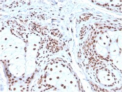
Biotium Nucleophosmin (Acute Myeloid Leukemia Marker)(NPM1/3286), CF568 conjugate, 0.1mg/mL
Recognizes a 33 kDa glycoprotein, identified as Nucleophosmin (NPM). It is predominantly localized in the nucleus of cells in most tissues. NPM is involved in ribosomal assembly and rRNA transport. It is an abundant protein that is highly phosphorylated by Cdc2 kinase during mitosis. This phosphoprotein moves between the nucleus and the cytoplasm. It is thought to be involved in several processes including regulation of the ARF/p53 pathway. A number of genes are fusion partners, in particular the anaplastic lymphoma kinase gene on chromosome 2. Mutations in exon 12 affecting the C-terminus of the protein are associated with an aberrant cytoplasmic location. Mutations in this gene are associated with acute myeloid leukemia. The antibody may be a useful aid for classification of acute myeloid leukemia. Primary antibodies are available purified, or with a selection of fluorescent CF Dyes and other labels. CF Dyes offer exceptional brightness and photostability. Note: Conjugat
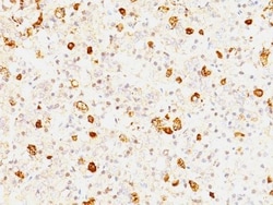
Biotium FSH beta(FSHb/1062), CF647 conjugate, 0.1mg/mL
This MAb reacts with a protein of 22 kDa, identified as beta sub-unit of FSH. It does not cross react with the alpha sub-unit. Follicle stimulating hormone (FSH) is a hormone synthesized and secreted by gonadotrophs in the anterior pituitary gland. In the ovary, FSH stimulates the growth of immature Graafian follicles to maturation. As the follicle grows, it releases inhibin, which deactivates the FSH production. In men, FSH enhances the production of androgen-binding protein by the Sertoli cells of the testis and is critical for spermatogenesis. FSH and LH act synergistically in reproduction. FSH is a useful marker in the classification of pituitary tumors and the study of pituitary disease.Primary antibodies are available purified, or with a selection of fluorescent CF Dyes and other labels. CF Dyes offer exceptional brightness and photostability. Note: Conjugates of blue fluorescent dyes like CF405S and CF405M are not recommended for detecting low abundance targets, b
Biotium NGF-Receptor (p75) / CD271 (Soft Tissue Tumor Marker) (NGFR/2550R), Biotin conjugate, 0.1mg/mL
This antibody recognizes a glycoprotein of 75 kDa, identified as low affinity Nerve Growth Factor (NGF) Receptor (p75NGFR) or Neurotrophin Receptor (p75NTR). NGFR is expressed in various neural crest cells and their tumors such as melanocytes, melanomas, neuroblastomas, pheochromocytomas and neurofibromas. Reportedly, anti-NGFR is a reliable marker for desmoplastic and neurotropic melanomas. NGFR is expressed in mature non-neural cells such as perivascular cells, dental pulp cells, lymphoidal follicular dendritic cells, basal epithelium of oral mucosa and hair follicles, prostate basal cells, and myoepithelial cells. Anti-NGFR stains the myoepithelial cells of breast ducts and intra-lobular fibroblasts of breast ducts.Primary antibodies are available purified, or with a selection of fluorescent CF Dyes and other labels. CF Dyes offer exceptional brightness and photostability. Note: Conjugates of blue fluorescent dyes like CF405S and CF405M are not recommended for detectin
Biotium CELA3B / ELA3B (Pancreatic Function Marker) (CELA3B/2809R), CF740 conjugate, 0.1mg/mL
This MAb recognizes a protein of ~30 kDa, identified as CELA3B (Chymotrypsin like elastase family member 3B). Elastases form a subfamily of serine proteases that hydrolyze many proteins in addition to elastin. Humans have six elastase genes which encode the structurally similar proteins elastase 1, 2, 2A, 2B, 3A, and 3B. Unlike other elastases, elastase 3B has little elastolytic activity. Like most of the human elastases, elastase 3B is secreted from the pancreas as a zymogen and, like other serine proteases such as trypsin, chymotrypsin and kallikrein; it has a digestive function in the intestine. Elastase 3B preferentially cleaves proteins after alanine residues. Elastase 3B may also function in the intestinal transport and metabolism of cholesterol. Both elastase 3A and elastase 3B have been referred to as protease E and as elastase 1, and excretion of this protein in fecal material is frequently used as a measure of pancreatic function.Primary antibodies are available purified
Biotium CELA3B / ELA3B (Pancreatic Function Marker) (CELA3B/1257), Biotin conjugate, 0.1mg/mL
This MAb recognizes a protein of ~30 kDa, identified as CELA3B (Chymotrypsin like elastase family member 3B). Elastases form a subfamily of serine proteases that hydrolyze many proteins in addition to elastin. Humans have six elastase genes which encode the structurally similar proteins elastase 1, 2, 2A, 2B, 3A, and 3B. Unlike other elastases, elastase 3B has little elastolytic activity. Like most of the human elastases, elastase 3B is secreted from the pancreas as a zymogen and, like other serine proteases such as trypsin, chymotrypsin and kallikrein; it has a digestive function in the intestine. Elastase 3B preferentially cleaves proteins after alanine residues. Elastase 3B may also function in the intestinal transport and metabolism of cholesterol. Both elastase 3A and elastase 3B have been referred to as protease E and as elastase 1, and excretion of this protein in fecal material is frequently used as a measure of pancreatic function in clinical assays.Primary antibodie
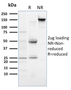
Biotium PLK1 (Marker of Mitosis)(AZ44), CF488A conjugate, 0.1mg/mL
Plk (for polo-like kinase) encodes a serine/threonine kinase that is closely related to polo and CDC5, genes that are required for passage through mitosis in Drosophila and Saccharomyces, respectively. Polo and Cdc5 both code for proteins that are involved in regulating the function of the mitotic spindle. Plk protein accumulates in the cell during the S and G2 phases of the cell cycle; Plk protein content and catalytic activity peak at the onset of mitosis, followed by a rapid reduction after mitosis. Plk expression is detectable in mitotically active tissues such as colon and placenta, as well as in tumors of various origins. It has also been suggested that Plk may serve as a marker of cell proliferation. Required for recovery after DNA damage checkpoint and entry into mitosis. Required for kinetochore localization of BUB1B. Phosphorylates SGOL1. Required for spindle pole localization of isoform 3 of SGOL1 and plays a role in regulating its centriole cohesion function. Phos
Biotium CD84(153-4D9), 1mg/mL
Recognizes a protein of 74 kDa, identified as CD84. It is expressed on mature B cells and on B-cell lines, including pre-B-cell lines, but not on plasma cell lines. Immunohistochemical studies demonstrated that CD84 strongly expressed on tissues macrophages. CD84 is also highly expressed on platelets and, at low levels on peripheral blood T cells. It is a highly N-glycosylated protein and belongs to immunoglobulin superfamily. It may play a role in leukocyte activation.Primary antibodies are available purified, or with a selection of fluorescent CF Dyes and other labels. CF Dyes offer exceptional brightness and photostability. Note: Conjugates of blue fluorescent dyes like CF405S and CF405M are not recommended for detecting low abundance targets, because blue dyes have lower fluorescence and can give higher non-specific background than other dye colors.
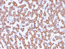
Biotium Prohibitin (Mitochondrial Marker)(PHB/3230), CF740 conjugate, 0.1mg/mL
Recognizes a protein of 30 kDa which is identified as Prohibitin, an evolutionarily conserved protein with homologues found in yeast to man. It is located in the inner membrane of mitochondria. Although prohibitin mRNA and protein expression occurs throughout the cell cycle, maximum levels are detected during the G1/S phase transition and minimum levels are seen in S phase and the G2/mitosis boundary. Prohibitin is located exclusively in the mitochondria with the highest concentration on the inner membrane. Prohibitin is an ideal mitochondrial marker. It shows antiproliferative activity and has been proposed to play a role in normal cell cycle regulation, replicative senescence, cellular immortalization, and tumor suppression. Primary antibodies are available purified, or with a selection of fluorescent CF Dyes and other labels. CF Dyes offer exceptional brightness and photostability. Note: Conjugates of blue fluorescent dyes like CF405S and CF405M are not recommended fo
Biotium CD11c (Dendritic Cell Marker)(ITGAX/1242), Biotin conjugate, 0.1mg/mL
Recognizes a protein of 145 kDa, identified as CD11c. CD11c (ITGAX), a member of the leukointegrin family, shares the same beta subunit with other members of the leukocyte adhesion molecule family, which includes CD11a (LFA-1), CD11b (MAC-1) and CD11d (ITGAD), but has a unique alpha chain. CD11c has been shown to play a role in phagocytosis, cell migration, and cytokine production by monocytes/macrophages as well as induction of T-cell proliferation by Langerhans cells. CD11c is expressed prominently on the plasma membranes of monocytes, tissue macrophages, NK cells, and most dendritic cells (DCs). A lower level of expression is also observed on neutrophils as a result of its high level of expression on most DCs. An antibody to CD11c may aid in identification of lesions with histiocytic origin. It may also been used as a marker for hairy cell leukemia in paraffin-embedded tissues.
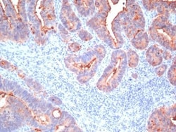
Biotium CD66, pan (CEA)(Cocktail), 0.2mg/mL
This antibody recognizes proteins of 80-200 kDa, identified as different members of CEA family. CEA is synthesized during development in the fetal gut and is re-expressed in increased amounts in intestinal carcinomas and several other tumors. This MAb does not react with nonspecific cross-reacting antigen (NCA) and with human polymorphonuclear leucocytes. It shows no reaction with a variety of normal tissues and is suitable for staining of formalin/paraffin tissues. CEA is not found in benign glands, stroma, or malignant prostatic cells. Antibody to CEA is useful in detecting early foci of gastric carcinoma and in distinguishing pulmonary adenocarcinomas (60-70% are CEA positive) from pleural mesotheliomas (rarely or weakly CEA positive). Anti-CEA positivity is seen in adenocarcinomas from the lung, colon, stomach, esophagus, pancreas, gallbadder, urachus, salivary gland, ovary, and endocervix.Primary antibodies are available purified, or with a selection of fluorescent CF
Biotium CD63 (Late Endosomes Marker)(NKI/C3 + LAMP3/968), 0.2mg/mL
This MAb recognizes protein of 26 kDa-60 kDa, which is identified as CD63. The tetraspanins are integral membrane proteins expressed on cell surface and granular membranes of hematopoietic cells and are components of multi-molecular complexes with specific integrins. The tetraspanin CD63 is a lysosomal membrane glycoprotein that translocates to the plasma membrane after platelet activation. CD63 is expressed on activated platelets, monocytes and macrophages, and is weakly expressed on granulocytes, T cell and B cells. It is located on the basophilic granule membranes and on the plasma membranes of lymphocytes and granulocytes. CD63 is a member of the TM4 superfamily of leukocyte glycoproteins that includes CD9, CD37 and CD53, which contain four transmembrane regions. CD63 may play a role in phagocytic and intracellular lysosome-phagosome fusion events. CD63 deficiency is associated with Hermansky-Pudlak syndrome and is strongly expressed during the early stages of melanoma pr
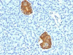
Biotium Insulin(E2-E3 + 2D11-H5), Biotin conjugate, 0.1mg/mL
Recognizes a polypeptide which is identified as insulin, a 51-amino acid polypeptide composed of A and B chains connected through the C-peptide. Proinsulin, which has very little biological activity, is cleaved by proteases within its cell of origin into the insulin molecule and the C-terminal basic residue. Insulin enhances membrane transport of glucose, amino acids, and certain ions. It also promotes glycogen storage, formation of triglycerides, and synthesis of proteins and nucleic acids. Deficiency of insulin results in diabetes mellitus. The main storage site for insulin is the pancreatic islets. Antibodies to insulin are important as beta-cell and insulinoma marker.Primary antibodies are available purified, or with a selection of fluorescent CF Dyes and other labels. CF Dyes offer exceptional brightness and photostability. Note: Conjugates of blue fluorescent dyes like CF405S and CF405M are not recommended for detecting low abundance targets, because blue dyes have
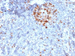
Biotium Cdk1 / p34cdc2 Serine-Threonine Kinase(A17.1.1), CF568 conjugate, 0.1mg/mL
Recognizes a 34 kDa protein (cdk1), identified as p34cdc2 (a catalytic subunit of Maturation Promoting Factor). Its epitope maps near the C-terminus of the protein and its core is thought to be LGTPNNEV (aa220-227 in murine cdc2). It shows no cross reaction with cdk2 p32. It supports the kinase activity. p34cdc2 plays a crucial role during cell division and is most active during mitosis. It is a serine/threonine kinase, which is activated by cyclin, by dephosphorylation of tyrosine residues. p34cdc2 is inactivated by a tyrosine kinase. This MAb reportedly inhibits the activation of p34cdc2 kinase by cyclins. Primary antibodies are available purified, or with a selection of fluorescent CF Dyes and other labels. CF Dyes offer exceptional brightness and photostability. Note: Conjugates of blue fluorescent dyes like CF405S and CF405M are not recommended for detecting low abundance targets, because blue dyes have lower fluorescence and can give higher non-specific background